Human Leg Bones Diagram / Long bones are found in the arms (humerus, ulna, radius) and legs (femur, tibia, fibula), as well as in.
byAdmin•
0
Human Leg Bones Diagram / Long bones are found in the arms (humerus, ulna, radius) and legs (femur, tibia, fibula), as well as in.. One is the ulna, and the other is the radius. Diagram depicting the arterial supply to a growing leg. The bones of the leg are the femur, tibia, fibula and patella.the foot bones shown in this diagram are the talus, navicular, cuneiform, cuboid, metatarsals and calcaneus. Human anatomy diagrams show internal organs, cells, systems, conditions, symptoms and sickness information and/or tips for healthy living. Each leg is made up of four bones.
Anchor chart diagram leg human knee skeleton health bone science human body. The patella (kneecap) is the sesamoid bone in front of the knee. This area is commonly referred to as the calf. The bones of the hip include the femur, the ilium, the ischium, and the pubis. The lower leg extends from the knee to the ankle.
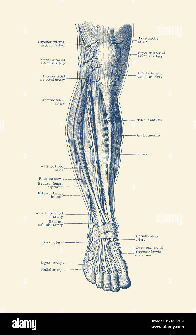
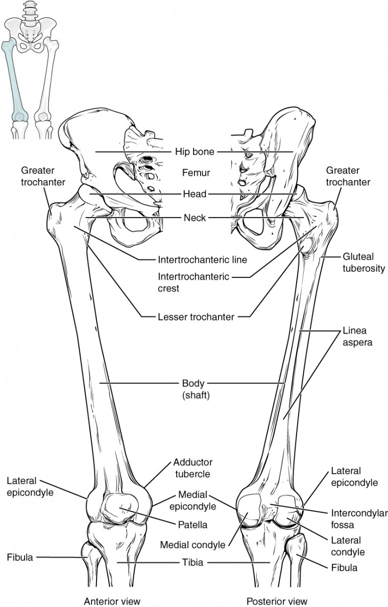
Vintage Anatomy Print Of The Human Leg Showcasing The Veins And Arteries Stock Photo Alamy from c8.alamy.com Its lower end helps create the knee joint. The thigh bone, or femur, is the large upper leg bone that connects the lower leg bones (knee joint) to the pelvic bone (hip joint). In concert with each other, the two bones play a vital role in how the forearm rotates. Long bones are found in the arms (humerus, ulna, radius) and legs (femur, tibia, fibula), as well as in. Health diagram bone skeleton leg knee science anchor chart human human body. The major bones of the leg are the femur (thigh bone), tibia (shin bone), and adjacent fibula, and these are all long bones. The hip itself is a ball and socket joint, much like the shoulder.the structures necessary to create this joint are the socket, the joint capsule, muscle, ligaments, and the neck. Each leg is made up of four bones.
Most of the leg skeleton has bony prominences and margins that can be palpated and some serve as anatomical landmarks that define the extent of the leg.
Tendons connect the knee bones to the leg muscles that move the knee. It is composed of 300 bones at birth, but later decreases to 80 bones in the axial skeleton and 126 bones in the appendicular skeleton. As these muscles contract and relax, they move skeletal bones to create movement of the body. One is the ulna, and the other is the radius. Health diagram bone skeleton leg knee science anchor chart human human body. The bones of the hip include the femur, the ilium, the ischium, and the pubis. A high ankle sprain causes pain and swelling similar to a. Start studying leg bone diagram. The patella (kneecap) is the sesamoid bone in front of the knee. The femur is the largest bone in the body and the only bone of the thigh (femoral) region. The lower leg extends from the knee to the ankle. The pubis, ischium, and ilium together constitute the pelvis while the thigh bone is the femur. These muscles work together to produce movements such as standing, walking, running, and jumping.
The pubis, ischium, and ilium together constitute the pelvis while the thigh bone is the femur. Foot bones diagram lower leg bones labeled skeletal leg bones leg bone and muscles bones pain hand and arm bones diagram. The femur, or thighbone, is the longest and largest bone in the human body. Diagramme schnell und einfach erstellen. The hip itself is a ball and socket joint, much like the shoulder.the structures necessary to create this joint are the socket, the joint capsule, muscle, ligaments, and the neck.
Thigh Anatomy Diagram Pictures Body Maps from post.greatist.com The femur is the largest bone in the body and the only bone of the thigh (femoral) region. The bones of the leg are the femur, tibia, fibula and patella. The femur or thighbone is the longest and largest bone in the human body. One is the ulna, and the other is the radius. Health diagram bone skeleton leg knee science anchor chart human human body. The thigh bone, or femur, is the large upper leg bone that connects the lower leg bones (knee joint) to the pelvic bone (hip joint). Start studying leg bone diagram. Posted on june 5, 2014 by admin.
The lower leg extends from the knee to the ankle.
Formed by the left and right hip bones, the pelvic girdle connects the lower limb (leg) bones to the axial skeleton. Human anatomy diagrams show internal organs, cells, systems, conditions, symptoms and sickness information and/or tips for healthy living. A high ankle sprain causes pain and swelling similar to a. The majority of muscles in the leg are considered long muscles, in that they stretch great distances. The bones of the hip include the femur, the ilium, the ischium, and the pubis. At the same time, the bones and joints of the leg and foot must be strong enough to support the body's weight while remaining. The foot bones shown in this diagram are the talus, navicular, cuneiform, cuboid, metatarsals and calcaneus. Health diagram bone skeleton leg knee science anchor chart human human body. The ligament joining the two bones of the lower leg (tibia and fibula), called the syndesmotic ligament, is injured. The hip itself is a ball and socket joint, much like the shoulder.the structures necessary to create this joint are the socket, the joint capsule, muscle, ligaments, and the neck. As these muscles contract and relax, they move skeletal bones to create movement of the body. This image is an edited version of this image that was created by user:ladyofhats (mariana ruiz villarreal). These bones are arranged into two major divisions:
These bones are arranged into two major divisions: These muscles work together to produce movements such as standing, walking, running, and jumping. Its lower end helps create the knee joint. Labeled human leg bones created for use in leg bone. In concert with each other, the two bones play a vital role in how the forearm rotates.
Bones Of The Lower Limb Anatomy And Physiology from s3-us-west-2.amazonaws.com In concert with each other, the two bones play a vital role in how the forearm rotates. The major bones of the leg are the femur (thigh bone), tibia (shin bone), and adjacent fibula, and these are all long bones. Each leg is made up of four bones. Side view of foot bones inter mediate gone gone gone talus gone ca can eug gone cuboid gone gores phalange go neg o 5th metatarsal gone The forearm contains two major bones. The ligament joining the two bones of the lower leg (tibia and fibula), called the syndesmotic ligament, is injured. The bones of the leg are the femur, tibia, fibula and patella.the foot bones shown in this diagram are the talus, navicular, cuneiform, cuboid, metatarsals and calcaneus. This area is commonly referred to as the calf.
The femur, or thigh bone, is the single bone of the thigh region (figure 6.51).
Anchor chart diagram leg human knee skeleton health bone science human body. See more ideas about muscle anatomy, human anatomy and physiology, body anatomy. The thigh bone, or femur, is the large upper leg bone that connects the lower leg bones (knee joint) to the pelvic bone (hip joint). Human body anatomy bones 12 photos of the human body anatomy bones human body bone flashcards, human body bones chest, human body bones definitions, human body bones tibia, human body. The smaller bone that runs alongside the tibia (fibula) and the kneecap (patella) are the other bones that make the knee joint. The forearm contains two major bones. The pubis, ischium, and ilium together constitute the pelvis while the thigh bone is the femur. Tendons connect the knee bones to the leg muscles that move the knee. The bones together make up the hip. Human skeleton anatomy human body anatomy human anatomy and physiology anatomy study anatomy drawing radiology student anatomy bones medical anatomy medical coding. The foot bones shown in this diagram are the talus, navicular, cuneiform, cuboid, metatarsals and calcaneus. Its lower end helps create the knee joint. The bones of the leg are the femur, tibia, fibula and patella.
Health diagram bone skeleton leg knee science anchor chart human human body leg bones diagram. The femur, or thighbone, is the longest and largest bone in the human body.